Researchers advance technique to detect ovarian cancer: Rice, MD Anderson use fluorescent carbon nanotube probes to achieve first in vivo success
Researchers at Rice University and the University of Texas MD Anderson Cancer Center have refined and, for the first time, run in vivo tests of a method that may allow nanotube-based probes to locate specific tumors in the body. Their ability to pinpoint tumors with submillimeter accuracy could eventually improve early detection and treatment of ovarian cancer.
The noninvasive technique relies on single-walled carbon nanotubes that can be optically triggered to emit shortwave infrared light.

Rice University and MD Anderson researchers have developed a technique that uses fluorescent nanotube-based probes to locate specific tumors in the body.
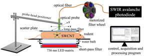
The Rice lab of chemist Bruce Weisman, a pioneer in the discovery and interpretation of the phenomenon, reported the new results recently that, the researchers used the technique to pinpoint small concentrations of nanotubes inside rodents. The lab of co-author Dr. Robert Bast Jr., an expert in ovarian cancer and vice president for translational research at MD Anderson, inserted gel-bound carbon nanotubes into the ovaries of rodents to mimic the accumulations that are expected for nanotubes linked to special antibodies that recognize tumor cells. The rodents were then scanned with the Rice lab's custom-built optical device to detect the faint emission signatures of as little as 100 picograms of nanotubes.
The device irradiated the rodents with intense red light from an array of light-emitting diodes and read fluorescent signals with a specialized sensitive detector. Because different types of tissue absorb emissions from the nanotubes differently, the scanner took readings from many locations to triangulate the tumor's exact location, as confirmed by later MRI scans.
Weisman said it should be possible to noninvasively find small ovarian tumors within rodents used for medical research by linking nanotubes to antibody biomarkers and administering the biomarkers intravenously. The biomarkers would accumulate at the tumor site. He said more refined versions of the optical scanner may then be able to locate a tumor within seconds, and further advances may extend the method’s application to human cancer detection. The new results suggested that antibody-nanotube probes could potentially detect tumors with as few as 100 ovarian cancer cells, which could make it a valuable tool for early detection.
Rice graduate student Ching-Wei Lin is lead author of the paper. Co-authors from the Bast group at MD Anderson are researcher Dr. Hailing Yang and senior research assistants Weiqun Mao and Lan Pang. Rice co-authors are chemistry graduate student Stephen Sanchez and Kathleen Beckingham, a professor of biosciences.
Source: Nanotechnology Now
- 549 reads
Human Rights
Fostering a More Humane World: The 28th Eurasian Economic Summi

Conscience, Hope, and Action: Keys to Global Peace and Sustainability

Ringing FOWPAL’s Peace Bell for the World:Nobel Peace Prize Laureates’ Visions and Actions

Protecting the World’s Cultural Diversity for a Sustainable Future

Puppet Show I International Friendship Day 2020

