Fluorescence Technique Measures Photoacid Distribution in Photoresists with Nanoscale Resolution
A team of researchers from the NIST Center for Nanoscale Science and Technology, the University of Maryland, and Korea University (Seoul, Korea) has measured the nanoscale distribution of photoacid molecules in photoresists using a fluorescence technique originally developed to provide images of biological structures smaller than the wavelength of light.* Photoresists are light-sensitive chemicals used for manufacturing the semiconductor integrated circuits found in computers and other electronics. By measuring the chemical reactions in photoresists at a smaller length scale, this method potentially opens a path to manufacturing smaller electronic devices.
In today's photoresists, chemical amplification of light allows for the use of low-brightness short-wavelength light sources, which enable smaller feature sizes to be printed at higher throughputs. Each individual photon activates a molecule which generates a photoacid. During post-exposure baking, the photoacid diffuses, rendering a volume of the resist soluble to developer. This improves the sensitivity, or photospeed, of the resist, but at the cost of degraded image resolution: a result of the photoacid diffusion. Until now, it has only been possible to infer indirectly where the photoacid molecules are produced or how they diffuse by analyzing the resist images after the resist is fully exposed, either before or after it is developed. A more direct measurement is critically needed, however, because the distribution of the photoacid molecules within a resist film limits the minimum feature sizes that can be produced on computer chips. Precise measurements can help photoresist manufacturers understand the processes that lead to loss of image contrast and develop measures to mitigate blur induced by photoacid diffusion.

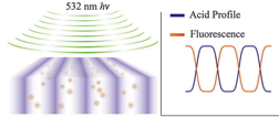
Schematic showing fluorescence from UV-activated fluorophores excited by 532 nm light that reveals nanoscale photoacid distribution (left). Activated fluorophore concentration corresponds to the inverse of the original photoacid distribution (right).
To observe the location of the photoacid molecules more directly, the research team used a novel fluorescent dye that can be switched from a dark to bright state either by exposure to ultraviolet light or by reaction with a nearby acid molecule. Over time, they fit the fluorescent signal of each dye molecule to a two-dimensional distribution, allowing them to map the locations of the associated photoacid molecules with single-molecule sensitivity. The team also developed new statistical analysis methods that enable them to extract high-resolution information even when there is a very low concentration of fluorescent molecules. This method allows them to be confident that the behavior of the system is not changed by the presence of the fluorophores.
Ultimately, the researchers believe that these techniques will be useful for measuring nanoscale transport processes in a wide variety of soft-matter systems beyond photoresists, such as in polymers.
Source: Nanotechnology Now
- 403 reads
Human Rights
Fostering a More Humane World: The 28th Eurasian Economic Summi

Conscience, Hope, and Action: Keys to Global Peace and Sustainability

Ringing FOWPAL’s Peace Bell for the World:Nobel Peace Prize Laureates’ Visions and Actions

Protecting the World’s Cultural Diversity for a Sustainable Future

Puppet Show I International Friendship Day 2020

